As the big day approaches to get ones braces on, there is usually a quick step that precedes that appointment. With modern technology, the newer braces can be made in small dots making them much more esthetic. These are bonded to the teeth by etching the enamel surface with acid. However, the molar teeth bear quite a bit of brunt of the bite, so bonding brackets onto those teeth can be frustrating because they can become debonded under normal chewing. Thus, many times it is helpful to go back to the old style bands for those molars. To seat the bands comfortably, there needs to be a small gap between the teeth. Therefore, separators must be placed ahead of time to create that space. A separator can be an elastic “donut” or a twisted wire. They are placed about a week before the big appointment. This will allow the bands to be slipped on with minimal fuss. They usually will make ones teeth sore for a day or two – nothing that a little ibuprofen wouldn’t take care of. But it is sure well worth it because otherwise, the bands would have to be forced on.
Category Archives: Dentistry
Making Room for Crowded Teeth
Crowded teeth are a very common problem affecting many people. To straighten these teeth orthodontically, a dentist needs to make room in the mouth to fit all of the teeth in a nice straight line. Many times, the crowding is so severe that teeth have to be removed to make room. However, it is preferable to keep all of the teeth (except for the wisdom teeth) if possible. This way, it is easier to get teeth to mesh properly. The other alternative is to reduce the size of the teeth.
Teeth are coated with enamel, the hard white substance that we see in our pearly whites. It has no nerve endings, just like hair and fingernails, so it can be smoothed and shaped without the need for local anesthetic. A quarter of a millimeter can be safely removed from each side in between the teeth without causing an increase in decay. That doesn’t sound like much, but when that small increment is added up over an entire arch of teeth, that can amount to a few millimeters which can be enough to allow for an adequate amount of space to makes ones teeth picket fence straight.
Periodontal Inflammation
Periodontal (or gum) inflammation is a response of the body to the onslaught of bacterial toxins and acids underneath the gums. Various factors such as diabetes, smoking, genetics, etc. can change the severity of the body’s response to these bacterial products. Inflammation results in redness and swelling of the gums. The body will send a lot of immune cells to try to remove the irritating substances that are residing in the gums. This immune response is essential for the body to ward off infections, however, in a situation where the oral hygiene is not good, the inflammatory response becomes chronic and the body is not able to return to normal health. This causes the eventual breakdown of the bone that holds the teeth in place. Symptoms of periodontal inflammation include bleeding and red gums. More advanced cases lead to loosening of the teeth and pus exuding from the gums.  The by-products of inflammation eventually overtake the body’s system of repair resulting in permanent damage. The only way to break this cycle is by performing meticulous oral hygiene (simple brushing and flossing) on a daily basis.
Sinus Lift Procedure for Dental Implants
To place an implant, there must be an adequate amount of bone to bear the brunt of the bite. In the upper molar area, it is very common to be short of bone for implants since the maxillary sinus is located right above it. Since the bone shrinks once a tooth is removed, it is not unusual to find only a few millimeters of bone remaining. In these cases, a procedure called a sinus lift bone graft is available.  In a sinus lift, a flap of gum is retracted from the upper jaw in the molar area, and a small window is cut into the bone being careful to not lacerate the sinus membrane. The sinus membrane is gently “lifted” and a bone graft material is placed in the hollowed out space in between the lower part of the sinus bone and the membrane. The surgical site is closed and allowed to heal undisturbed for a number of months. At this point, the bone will be fully organized and ready for implant placement. A sinus lift procedure has over 90% success rate. The feedback that I have gotten from patients as far as the recovery from the procedure has been very favorable. The technique opens up many more opportunities to restore the upper jaw with implants instead of having to resort to dentures.
Bone Augmentation for Implants
For an implant to be successful, there must be adequate bone available for proper placement. Since the bone and gums will shrink away once a tooth is removed, it is not unusual to have a situation where there is an inadequate amount of bone to place an implant. Simple techniques start with socket preservation where the dentist places a bone grafting material in the socket at the time of tooth removal. Even more predictable is placing a membrane over the graft material. This will allow the graft to heal undisturbed and has a very high rate of success. Once the bone height has shrunk, then a block graft needs to be done to give the bone more vertical. A block graft uses a piece of cortical bone that is obtained from another part of the body. Common areas are the mandibular ramus (the part of the jaw bone that lies behind the lower back teeth) or the mental symphysis (part of the chin). Once a block of bone has been harvested, then a flap of gum is peeled back at the donor site and the block is held in place with screws. Since the procedure utilizes bone from the same person, it doesn’t reject the graft. After allowing the area to heal for a few months, then the site will be ready for implant placement.
Bone Augmentation for Dentures (Part II of II)
There are a number of techniques available to build up the jaw bone to give a good base for dentures. With autogenous bone grafts, a piece of bone is harvested from another part of the patient’s body. The most common area to get the bone from is the iliac crest. This is the part of the pelvis that lies above the hip joint. The body tolerates the bone graft since it is from the same person, so there is no risk for rejection. However, the surgical procedure that is utilized to obtain the bone can be somewhat debilitating. The feedback that I have gotten from patients is that the hip was much more uncomfortable than the mouth. Another technique is an allograft. This procedure utilizes cadaver bone. The sterilizing and screening techniques make for a wide margin of safety for cadaver bone. And the great news is there is not a need for an additional painful surgical site and long recovery period. Although the surgical success rate is good with allografts, they are still not as predictable as autogenous grafts (utilizing the body’s own tissue). After a few months of healing time, a denture can be safely constructed that should provide years of service.
Bone Augmentation for Dentures (Part I of II)
To place dentures, there must be an adequate amount of jaw bone available to provide a place for the dentures to seat. Soft tissue is not firm enough to give proper support for false teeth. Alveolar bone is the bone that holds the teeth in place. That is its only purpose, so once the teeth have been removed, the body figures it has no more use for it and it resorbs (dissolves away).  If the teeth have been missing for too long, then the available bone to hold a denture on is virtually nonexistent. That’s where bone augmentation helps. It is a surgical procedure to add bone to selected areas of the mouth. This will increase the height of the bony ridge and will give the denture more stability. There are a number of techniques available to add height to the jaw bone. These are particulate and bone graft substitutes. There are fancier membrane techniques where a thin membrane covers the graft material and allows the graft to heal undisturbed. There are autogenous bone grafts where a piece of bone form one’s own body is used and there are allografts which utilize cadaver bone for its source. The ridges can also be widened utilizing distraction osteogenesis. I will discuss each technique in more detail in Part II.
X-rays in Dentistry (Part II of II)
X-rays are extremely important in dentistry to provide early detection of cavities and gum disease. However, there are many other uses. Since x-rays provide an image of what is inside the body, it can be used to see tumors and cysts that would otherwise go undetected. Early detection of tumors can be life-saving. Even though cysts, which are fluid-filled sacs, are not cancerous, they can expand and cause damage to bone. By periodically x-raying cysts, your dentist can see if it is beginning to grow and time surgical removal appropriately.
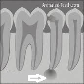
X-rays can also be used to detect dead nerves in teeth. Although x-rays cannot provide an image of soft tissue, once the dead nerve has caused damage to the bone surrounding the apex, or tip, of the root, it can be spotted on an x-ray film. X-rays can also be used in orthodontics to relate the upper and lower jaws to the rest of the skull. This can give an important insight on how to proceed with treatment. They can also be used to track growth and development in children. Another use of x-rays is to view implant placement to make sure that it is lined up properly during the surgery. They can also be helpful in checking the fit of crown margins. It is very important that the margin, or edge, of a crown meet the tooth surface perfectly. X-rays can see whether the margin is too short, too long, or open. Since these margins are usually tucked under the gums, x-rays provide a very useful adjunct to proper placement. As you can see, x-rays in dentistry are not just for checking cavities.
X-rays in Dentistry (Part I of II)
X-rays were discovered in 1895 by German physicist, Wilhelm Röntgen. He also discovered the medical application of x-rays when he passed his hand in front of a barium screen and noticed a shadow of his skeleton. The ability to see inside the body has been a big boom for diagnosis in dentistry. A tiny burst of x-rays aimed between the teeth will show cavities in their early stages before they can be detected by visual examination. As the x-rays pass through the teeth, the high density of tooth structure will stop the x-rays from reaching the film. Decay is less dense because the acid from bacterial plaque leaches out the calcium. Therefore, more x-rays are able to pass through that point. The more x-rays that pass through, the darker the point on the x-ray film. The dentist will look at the film for dark spots against a white background in order to detect decay. The middle and later stages of gum disease can also be detected because the disease will attack the bone. With less density of the bone, the damaged bone will show up on an x-ray film as darker and therefore the extent of the disease can be better quantified.
Bleeding Gums
If you ever notice blood on your tooth brush after brushing your teeth, chances are that there is gum disease present. There should not be one drop of blood present on a brush or floss after use. These warning signs should be heeded earlier as opposed with later. Before you run to the dentist with bleeding gums, it is best to try a regimen of good oral hygiene first. Just simple but thorough brushing and flossing a couple of times a day can go a long way in stopping bleeding gums. I have actually had patients who have told me that they purposely did not floss because every time that they did, it made their gums bleed. What they didn’t realize was that by not flossing, it left the bacterial plaque behind that is the cause of the bleeding in the first place. The patients were thinking that the mechanical cleaning with the floss was what was causing the bleeding to occur, whereas, it was actually the gum disease itself that caused the problem. It may still take a trip to the dentist for a professional cleaning for the bleeding to subside. But by doing the good home care ahead of time, it can be the first step toward eliminating gum disease in your mouth.


