To place dentures, there must be an adequate amount of jaw bone available to provide a place for the dentures to seat. Soft tissue is not firm enough to give proper support for false teeth. Alveolar bone is the bone that holds the teeth in place. That is its only purpose, so once the teeth have been removed, the body figures it has no more use for it and it resorbs (dissolves away).  If the teeth have been missing for too long, then the available bone to hold a denture on is virtually nonexistent. That’s where bone augmentation helps. It is a surgical procedure to add bone to selected areas of the mouth. This will increase the height of the bony ridge and will give the denture more stability. There are a number of techniques available to add height to the jaw bone. These are particulate and bone graft substitutes. There are fancier membrane techniques where a thin membrane covers the graft material and allows the graft to heal undisturbed. There are autogenous bone grafts where a piece of bone form one’s own body is used and there are allografts which utilize cadaver bone for its source. The ridges can also be widened utilizing distraction osteogenesis. I will discuss each technique in more detail in Part II.
Monthly Archives: September 2014
X-rays in Dentistry (Part II of II)
X-rays are extremely important in dentistry to provide early detection of cavities and gum disease. However, there are many other uses. Since x-rays provide an image of what is inside the body, it can be used to see tumors and cysts that would otherwise go undetected. Early detection of tumors can be life-saving. Even though cysts, which are fluid-filled sacs, are not cancerous, they can expand and cause damage to bone. By periodically x-raying cysts, your dentist can see if it is beginning to grow and time surgical removal appropriately.
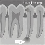
X-rays can also be used to detect dead nerves in teeth. Although x-rays cannot provide an image of soft tissue, once the dead nerve has caused damage to the bone surrounding the apex, or tip, of the root, it can be spotted on an x-ray film. X-rays can also be used in orthodontics to relate the upper and lower jaws to the rest of the skull. This can give an important insight on how to proceed with treatment. They can also be used to track growth and development in children. Another use of x-rays is to view implant placement to make sure that it is lined up properly during the surgery. They can also be helpful in checking the fit of crown margins. It is very important that the margin, or edge, of a crown meet the tooth surface perfectly. X-rays can see whether the margin is too short, too long, or open. Since these margins are usually tucked under the gums, x-rays provide a very useful adjunct to proper placement. As you can see, x-rays in dentistry are not just for checking cavities.

