X-rays in Dentistry (Part II of II)

X-rays are extremely important in dentistry to provide early detection of cavities and gum disease. However, there are many other uses. Since x-rays provide an image of what is inside the body, it can be used to see tumors and cysts that would otherwise go undetected. Early detection of tumors can be life-saving. Even though cysts, which are fluid-filled sacs, are not cancerous, they can expand and cause damage to bone. By periodically x-raying cysts, your dentist can see if it is beginning to grow and time surgical removal appropriately.

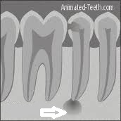
X-ray shows damage to bone at apex of root

X-rays can also be used to detect dead nerves in teeth. Although x-rays cannot provide an image of soft tissue, once the dead nerve has caused damage to the bone surrounding the apex, or tip, of the root, it can be spotted on an x-ray film. X-rays can also be used in orthodontics to relate the upper and lower jaws to the rest of the skull. This can give an important insight on how to proceed with treatment. They can also be used to track growth and development in children. Another use of x-rays is to view implant placement to make sure that it is lined up properly during the surgery. They can also be helpful in checking the fit of crown margins. It is very important that the margin, or edge, of a crown meet the tooth surface perfectly. X-rays can see whether the margin is too short, too long, or open. Since these margins are usually tucked under the gums, x-rays provide a very useful adjunct to proper placement. As you can see, x-rays in dentistry are not just for checking cavities.

X-rays in Dentistry (Part I of II)

Panoramic (full-mouth) x-ray

X-rays were discovered in 1895 by German physicist, Wilhelm Röntgen. He also discovered the medical application of x-rays when he passed his hand in front of a barium screen and noticed a shadow of his skeleton. The ability to see inside the body has been a big boom for diagnosis in dentistry. A tiny burst of x-rays aimed between the teeth will show cavities in their early stages before they can be detected by visual examination. As the x-rays pass through the teeth, the high density of tooth structure will stop the x-rays from reaching the film. Decay is less dense because the acid from bacterial plaque leaches out the calcium. Therefore, more x-rays are able to pass through that point. The more x-rays that pass through, the darker the point on the x-ray film. The dentist will look at the film for dark spots against a white background in order to detect decay. The middle and later stages of gum disease can also be detected because the disease will attack the bone. With less density of the bone, the damaged bone will show up on an x-ray film as darker and therefore the extent of the disease can be better quantified.

Bitewing X-rays

Bitewing x-rays are the routine x-rays that the dentist takes to check for cavities in between the teeth. These x-rays are important because tiny cavities can be spotted when they first start forming. Catching cavities at that stage is extremely helpful in catching decay while it is small and easily treated with just a filling. If a cavity gets large enough that it can be seen with the naked eye then there is a great possibility that the decay could already be in the nerve. If that is the case, then what could have been a few hundred dollars worth of dentistry to fix could suddenly cost thousands of dollars to repair. Bitewing x-rays are also very helpful in seeing bone damage due to periodontal (gum) disease. As the disease progresses, it causes the bone that holds the teeth in to erode away. If left unchecked, the damage to the bone could be so severe that teeth could be lost. Seeing this damage in it’s early stages can alert the dentist that there is a problem brewing. Since once bone is lost, the damage is irreversible, so catching bone loss before before it happens is very important.

Digital Dental X-Rays

digital dental x-rayWe finally added digital x-rays to our office. It was a big expense but it was well worth it. Digital x-rays have been available for a number of years, however, the quality didn’t come close to film. With the recent advances in technology, the number of megapixels available now can match good quality x-ray film. There are two types of digital x-rays. One system uses a phosphor plate in place of the film, the other uses a direct sensor just like the light sensor in your digital camera. The phosphor plate must be sent through a scanner which is then processed by a computer which inherently causes a delay in viewing the resulting radiograph. digital x-ray sensorsThe sensor type provides almost instantaneous feedback due to to it’s direct connection to the computer, however, the sensor is quite bulky and can be uncomfortable. We chose the phosphor plate system due to it’s sensor (it’s thinner than regular x-ray film) for the ultimate comfort for our patients. The radiation dosage that is needed to obtain good radiographs is less than that of film and there is no messy chemicals to fool with during processing.

Periapical X-Rays

Sample of a Periapical X-RayA periapical x-ray is a closeup of a particular tooth or teeth. If your dentist recommends one, by all means, have him take it. I recommend periapical x-rays to my patients anytime they say that they are having pain in a particular area, if there is deep decay in a tooth or if there is a problem or defect that is apparent on a bitewing x-ray (the x-ray that is used to detect cavities and gum disease) but cannot be seen in it’s entirety. When viewing a periapical x-ray, the dentist will look at the bone around the root, the height of the bone for periodontal (gum) defects, intactness of the root and for any other pathology (disease process) that could be visible. If a darker area exists on the x-ray around the tip of the root, then that could mean that there is a dead nerve inside the tooth. Dark areas up the side of the root could mean a possible root fracture. Dark areas at the crest of the bone (the portion closest to the crown of the tooth, i.e. the part of the tooth that you see) is usually an indication of bone loss due to gum disease. Pathological conditions can show up as either light or dark areas. This could be indicative of a tumor or cyst. That’s why it is so important to allow your dentist to take a periapical x-ray if he recommends it.

3D Dental X-Rays

3D Cone Beam X-RayWe now have a piece of “Star Wars” technology at our disposal. 3D x-rays are now readily available to dentists. Called cone beam tomography, one can see jaw structures, tooth relationships and root canal anatomy. They are becoming the standard of care for implant surgery since it shows the relationship between the available bone and major structures such as nerves and sinuses. It’s indispensable for making sure that a dental implant does not impinge on the nerve that controls the lower lip. It can also help find some hidden bone around the sinuses allowing for placement without doing expensive bone grafts. In orthodontics, the relationship between the upper and lower jaw in conjunction with the rest of the skull can be determined readily with these x-rays. The3D view can aid a dentist in search for an elusive root canal or an extremely curved root.

Although the amount of radiation needed to produce these x-rays is reasonably minimal, the general feeling at this time is to not make them a routine procedure, especially in children who will probably be exposed to multiple sources of radiation throughout their lifetime.


Panoramic X-Rays

Panoramic X-Ray MachinePantomographic radiographs, panoramic x-rays for short, show a picture of the entire upper and lower jaws and the surrounding structures. They are an excellent tool for your dentist to find dental diseases in there early stages. One can see tumors while they are small and have caused minimal damage. Removal at that stage can be less debilitating, and, if cancerous, can greatly increase the survival rate. One can see impacted wisdom teeth and their relationship to major structures such as nerves and sinuses. They are great for an orthodontist to check root alignment and blunting of the roots The detail is not enough to spot cavities in their early stages. It is also not generally used to diagnose gum disease since the closeup x-rays give better detail.

The amount of radiation is about the equivalent of a chest x-ray. It is not recommended on a routine basis, except for every third year. It can be an excellent screening tool to see things that don’t normally show up on the routine x-rays such as failing root canals, root fractures, or cysts. The use of panoramic x-rays can help keep costs of dental treatment down due to being able to catch things in their early stages and can also save lives.